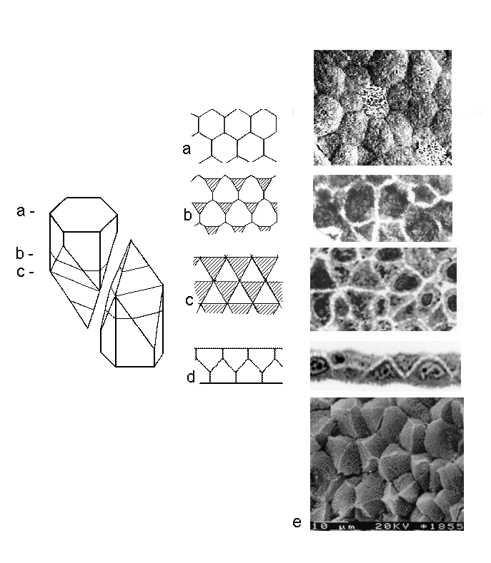
The first model of three-dimensional organization of a two-layered epithelia consisting of AB hexagonal cells. Left part of the Fig. shows a shape and mutual arrangement of cells. The superior and inferior parts of the model have the same construction but opposite orientation. The (a-c) part in the middle of the Fig. is the mosaic, corresponding to the tangential sections. It demonstrates the neighbouring cells at the different levels of the layer. The cellular shape of apicobasal section (across the cellular centers) of the model is presented on the scheme (d). Right part shows a real pattern of cell connections at the corresponding levels. From up to down it is shown:
Back to the Reconstruction of 3-D epithelial architecture
Back to the Picture Gallery
Copyright © 2000. All rights reserved
Back to the Homepage