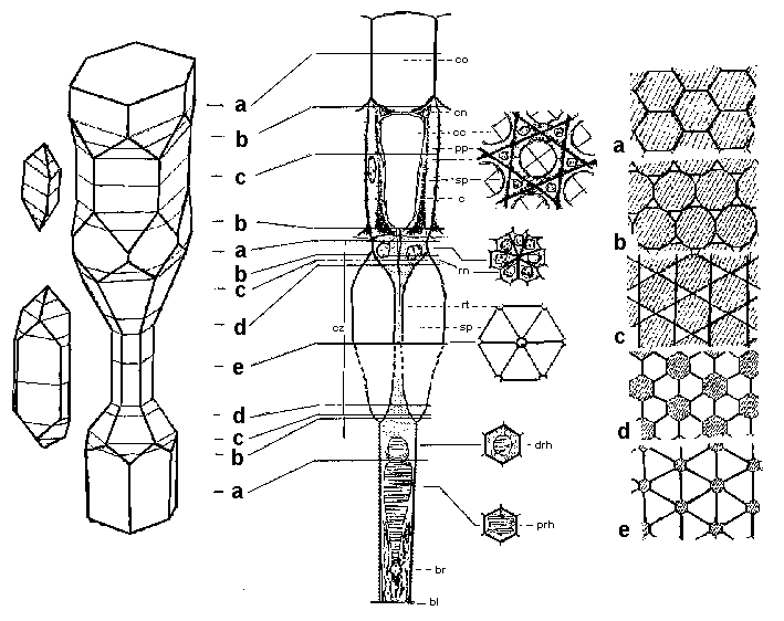

The simple 3-D model of the Scarabaeidae apposition eye consisting of opto-sensory complex (A) and secondary pigment cells (B) in ratio AB2. From left - the model of shape and interposition of eye elements; in the middle - schematic representation of real eye structural elements (from Caveney 1998, modificated); in right - the set of a - e mosaics that represents the eye elements contiguity on the various levels. The letters between model and scheme are indicate the levels where we can see the corresponding contiguity of eye elements.
bl, basal lamina; br, basal retinular cell; c, cone cell; cc, crystalline cone; cn, cone cell nucleus; co, corneal facet; cz, clear zone; drh, distal rhabdome; pp, peimary pigment cell; prh, proximal rhabdome; rn, retinular cell nucleus; rt, retinular cell tract; sp, secondary pigment cell. Montage from Savosryanov 2005.
Back to the Reconstruction of 3-D epithelial architecture
Back to the Picture Gallery
Back to the Homepage