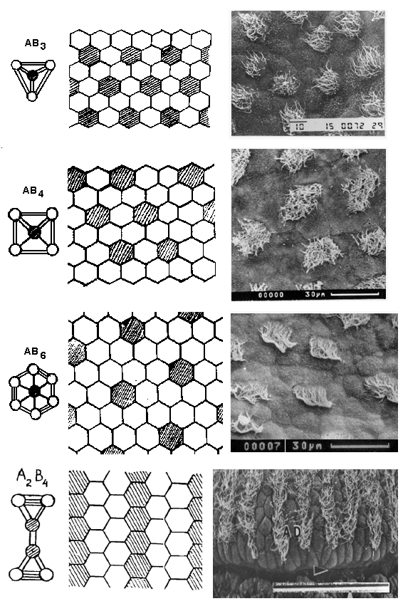
Models of ETU (left) and 2-D cell mosaics consisting of two cell types (A & B), characterized with a variable cell composition of the 1/3, 1/4 and 1/6 ratio. A real pattern of cell connections in grass frog larva epidermis is given at the right side. At the foot of the page there are the model and real cell mosaic, consisting of villous and smooth cells, appearing as alteration of strips in Cheilostomata ascophora larva epidermis. The last photo modified from Zimmer a. Woollacott, 1989. SEM. Composition from Savostyanov 2005.
Back to the Reconstruction of 3-D epithelial architecture
Back to the Picture Gallery
Back to the Homepage